OPS
OpenPolScope
Discover the Order in Living Things
Home
About us
Team
Contact
Locations
Collaborators
Technology
Overview
FAQ
- Birefringence
- Polarized Fluorescence
- Diattenuation
Literature
OPS Resource
Overview
Hardware
Services
Information Request Form
Software
Overview
Downloads
Documentation
Research
Overview
Polarized Light Field
MultiFocus PolScope
Tutorials
Overview
Color Legends
Universal Polarizer
Topics in Optics
- Hertzian Dipole
- Polarized Light
- Calcite Crystal Ellipsoid
Glossary
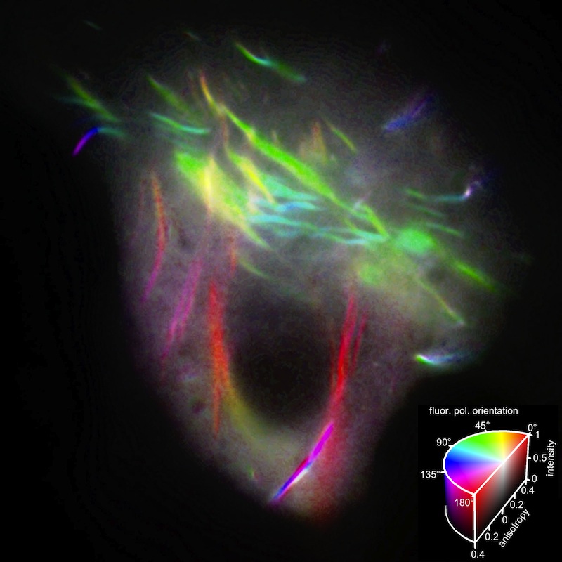
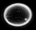
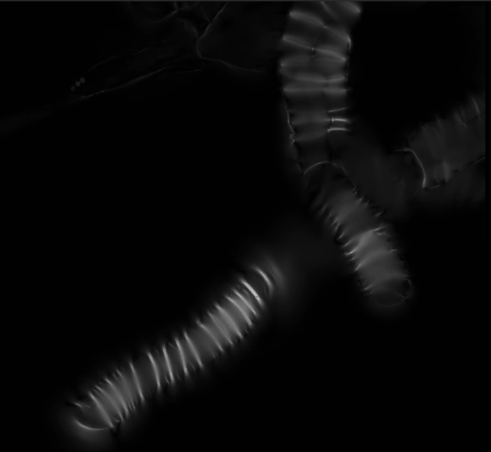
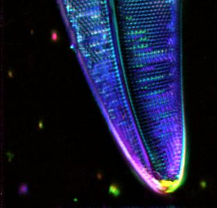
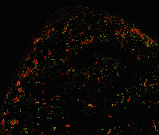
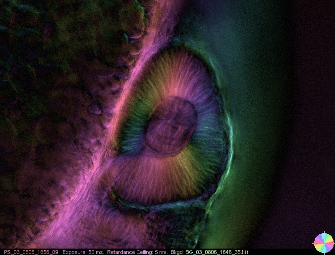
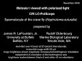
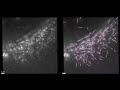
Gallery
Gallery
External Gallery
Gallery
© OpenPolScope | All Rights Reserved |
Contact
Last Page Update on June 12 2024 15:39